|
Cell Signaling Technology Inc
anti p her2 Anti P Her2, supplied by Cell Signaling Technology Inc, used in various techniques. Bioz Stars score: 94/100, based on 1 PubMed citations. ZERO BIAS - scores, article reviews, protocol conditions and more https://www.bioz.com/product/anti+p+her2/Phospho-HER2%2FErbB2+(Tyr1248)%2FEGFR+(Tyr1173)+Antibody/pmc12904715-217-20-21 Average 94 stars, based on 1 article reviews
anti p her2 - by Bioz Stars,
2026-10
94/100 stars
|
Buy from Supplier |
|
Danaher Inc
anti p erbb2 ab47755 Anti P Erbb2 Ab47755, supplied by Danaher Inc, used in various techniques. Bioz Stars score: 99/100, based on 1 PubMed citations. ZERO BIAS - scores, article reviews, protocol conditions and more https://www.bioz.com/product/anti+p+her2/Anti-ErbB2+%2F+HER2+antibody/pm24818668-53-4-9 Average 99 stars, based on 1 article reviews
anti p erbb2 ab47755 - by Bioz Stars,
2026-10
99/100 stars
|
Buy from Supplier |
|
Upstate Biotechnology Inc
p tyr1248 her 2  P Tyr1248 Her 2, supplied by Upstate Biotechnology Inc, used in various techniques. Bioz Stars score: 86/100, based on 1 PubMed citations. ZERO BIAS - scores, article reviews, protocol conditions and more https://www.bioz.com/product/anti+p+her2/anti+igg+rabbit/10__1158_slash_0008___5472__can___04___3841-52-86-89 Average 86 stars, based on 1 article reviews
p tyr1248 her 2 - by Bioz Stars,
2026-10
86/100 stars
|
Buy from Supplier |
|
Cell Signaling Technology Inc
anti p erbb2 Anti P Erbb2, supplied by Cell Signaling Technology Inc, used in various techniques. Bioz Stars score: 94/100, based on 1 PubMed citations. ZERO BIAS - scores, article reviews, protocol conditions and more https://www.bioz.com/product/anti+p+her2/Phospho-HER2%2FErbB2+(Tyr1221%2F1222)+Antibody/pm39094301-52-29-31 Average 94 stars, based on 1 article reviews
anti p erbb2 - by Bioz Stars,
2026-10
94/100 stars
|
Buy from Supplier |
|
Cell Signaling Technology Inc
anti p erbb2 tyr1221 1222 Anti P Erbb2 Tyr1221 1222, supplied by Cell Signaling Technology Inc, used in various techniques. Bioz Stars score: 96/100, based on 1 PubMed citations. ZERO BIAS - scores, article reviews, protocol conditions and more https://www.bioz.com/product/anti+p+her2/Phospho-HER2%2FErbB2+(Tyr1221%2F1222)+Rabbit+mAb/pm31059070-61-152-160 Average 96 stars, based on 1 article reviews
anti p erbb2 tyr1221 1222 - by Bioz Stars,
2026-10
96/100 stars
|
Buy from Supplier |
|
Cell Signaling Technology Inc
rabbit anti p erbb2 tyr1248  Rabbit Anti P Erbb2 Tyr1248, supplied by Cell Signaling Technology Inc, used in various techniques. Bioz Stars score: 94/100, based on 1 PubMed citations. ZERO BIAS - scores, article reviews, protocol conditions and more https://www.bioz.com/product/anti+p+her2/Phospho-HER2%2FErbB2+(Tyr1248)+Antibody/pmc05663201-15-0-7 Average 94 stars, based on 1 article reviews
rabbit anti p erbb2 tyr1248 - by Bioz Stars,
2026-10
94/100 stars
|
Buy from Supplier |
|
Cell Signaling Technology Inc
rabbit anti p her2 tyr 1248  Rabbit Anti P Her2 Tyr 1248, supplied by Cell Signaling Technology Inc, used in various techniques. Bioz Stars score: 94/100, based on 1 PubMed citations. ZERO BIAS - scores, article reviews, protocol conditions and more https://www.bioz.com/product/anti+p+her2/MELK+Antibody/bio_rxiv__2025__03__09__642136-42-28-33 Average 94 stars, based on 1 article reviews
rabbit anti p her2 tyr 1248 - by Bioz Stars,
2026-10
94/100 stars
|
Buy from Supplier |
|
Santa Cruz Biotechnology
anti p her2 Anti P Her2, supplied by Santa Cruz Biotechnology, used in various techniques. Bioz Stars score: 93/100, based on 1 PubMed citations. ZERO BIAS - scores, article reviews, protocol conditions and more https://www.bioz.com/product/anti+p+her2/p-Neu+Antibody/pmc09662874-54-12-18 Average 93 stars, based on 1 article reviews
anti p her2 - by Bioz Stars,
2026-10
93/100 stars
|
Buy from Supplier |
|
Upstate Biotechnology Inc
p erbb2  P Erbb2, supplied by Upstate Biotechnology Inc, used in various techniques. Bioz Stars score: 86/100, based on 1 PubMed citations. ZERO BIAS - scores, article reviews, protocol conditions and more https://www.bioz.com/product/anti+p+her2/anti+erbb2+p/10__1074_slash_jbc__m703811200-58-6-9 Average 86 stars, based on 1 article reviews
p erbb2 - by Bioz Stars,
2026-10
86/100 stars
|
Buy from Supplier |
|
Ayerst Laboratories
sirolimus Sirolimus, supplied by Ayerst Laboratories, used in various techniques. Bioz Stars score: 90/100, based on 1 PubMed citations. ZERO BIAS - scores, article reviews, protocol conditions and more https://www.bioz.com/product/anti+p+her2/sirolimus/pm15334195-54-39-55 Average 90 stars, based on 1 article reviews
sirolimus - by Bioz Stars,
2026-10
90/100 stars
|
Buy from Supplier |
|
Cell Signaling Technology Inc
rabbit monoclonal anti p erbb2 Rabbit Monoclonal Anti P Erbb2, supplied by Cell Signaling Technology Inc, used in various techniques. Bioz Stars score: 95/100, based on 1 PubMed citations. ZERO BIAS - scores, article reviews, protocol conditions and more https://www.bioz.com/product/anti+p+her2/HER2%2FErbB2+Rabbit+mAb/pm25099161-63-5-38 Average 95 stars, based on 1 article reviews
rabbit monoclonal anti p erbb2 - by Bioz Stars,
2026-10
95/100 stars
|
Buy from Supplier |
Image Search Results
Journal: Cancer Research
Article Title: Insulin-like Growth Factor-I Receptor/Human Epidermal Growth Factor Receptor 2 Heterodimerization Contributes to Trastuzumab Resistance of Breast Cancer Cells
doi: 10.1158/0008-5472.can-04-3841
Figure Lengend Snippet: Figure 2. Evidence of receptor cross talk: IGF-I induces phosphorylation of HER-2 in trastuzumab-resistant cells. A, parental SKBR3 cells were serum starved overnight and then stimulated with IGF-I (100 ng/mL) for 0, 1, 5, 10, or 15 minutes. Protein lysates (50 Ag) were examined by immunoblotting for p-IGF-IR (Tyr1163), total IGF-IR, p-HER-2 (Tyr1248), total HER-2, and actin. IGF-I stimulated phosphorylation of IGF-IR within 5 minutes. HER-2 phosphorylation was not significantly altered by IGF-I stimulation in parental cells. In addition, downstream signaling was studied by immunoblotting protein lysates (50 Ag) for p-IRS-1 (Ser789), total IRS-1, p-Akt (Ser473), total Akt, p-MAPK (Thr202/Tyr204 p42/p44 MAPK), total MAPK (p42/p44), p27kip1, and actin. Within 10 minutes of IGF-I stimulation, downstream signaling was activated with increased phosphorylation of IRS-1, Akt, and MAPK and down-regulation of p27kip1. B, resistant pool 2 cells were serum starved overnight and then stimulated with IGF-I (100 ng/mL) for 0, 1, 5, 10, 15, or 20 minutes. Protein lysates (50 Ag) were examined by immunoblotting for p-IGF-IR (Tyr1163), total IGF-IR, p-HER-2 (Tyr1248), total HER-2, and actin. Rapid phosphorylation of IGF-IR was evident within 1 minute. Importantly, IGF-I stimulated phosphorylation of HER-2 in resistant cells within 15 minutes. Downstream signaling was studied by immunoblotting protein lysates (50 Ag) for p-IRS-1 (Ser789), total IRS-1, p-Akt (Ser473), total Akt, p-MAPK (Thr202/Tyr204 p42/p44 MAPK), total MAPK (p42/p44), p27kip1, and actin. Within 1 minute of IGF-I stimulation, downstream signaling was activated with increased phosphorylation of IRS-1, Akt, and MAPK and down-regulation of p27kip1. C, parental and resistant pool 2 cells were serum starved overnight and then stimulated with IGF-I (100 ng/mL) for 0, 5, 10, or 15 minutes. IGF-IR was immunoprecipitated (200 Ag) and immunoblotted for HER-2 or IGF-IR. IGF-I stimulation did not affect the HER-2/IGF-IR heterodimer in resistant cells and seemed to promote interaction in parental cells.
Article Snippet: Total protein extracts (50 Ag) were immunoblotted using the following antibodies at the indicated dilutions: polyclonal antibodies for EGF receptor (EGFR), integrin h3, and platelet-derived growth factor receptor (PDGFR) at 1:1000 (Santa Cruz Biotechnology, Santa Cruz, CA); polyclonal p-Tyr1162/ 1163 IR/IGF-IR at 1:500 (BioSource); HER-2 monoclonal Ab-3 at 1:1,000 (Oncogene Research Products); p-Ser473-Akt (monoclonal 587F11) and polyclonal antibodies against IGF-IR, insulin receptor substrate 1 (IRS-1), Akt, p-Thr202/Tyr204 p42/p44 mitogen-activated protein kinase (MAPK), and total p42/p44 MAPK at 1:1,000 (Cell Signaling); polyclonal against p-Ser789-IRS-1 and polyclonal against
Techniques: Phospho-proteomics, Western Blot, Immunoprecipitation

Journal: Cancer Research
Article Title: Insulin-like Growth Factor-I Receptor/Human Epidermal Growth Factor Receptor 2 Heterodimerization Contributes to Trastuzumab Resistance of Breast Cancer Cells
doi: 10.1158/0008-5472.can-04-3841
Figure Lengend Snippet: Figure 5. Blockade of HER-2 by anti-HER-2 monoclonal antibody pertuzumab partially disrupts IGF-IR/HER-2 dimerization and slightly decreases viability of resistant cells. SKBR3-derived resistant cells were treated for 72 hours with 0, 1, or 10 Ag/mL of pertuzumab. A, IGF-IR was immunoprecipitated from protein lysates (200 Ag) and immunoblotted for HER-2 or IGF-IR. HER-2/IGF-IR heterodimerization was partially disrupted in cells treated with pertuzumab. B, total lysates (50 Ag) were immunoblotted for p-Tyr1248 HER-2, total HER-2, p-Tyr1163-IGF-IR, and total IGF-IR. Pertuzumab treatment did not affect phosphorylation or total levels of IGF-IR or HER-2. C, total lysates (50 Ag) were immunoblotted for p-IRS-1 (Ser789), total IRS-1, p-Akt (Ser473), total Akt, p-MAPK (Thr202/Tyr204 p42/p44 MAPK), total MAPK (p42/p44), p27kip1, and actin. Signaling to Akt was partially inhibited and p27kip1 was induced. D, parental cells and pools were treated in triplicate with 2-fold serial dilutions of pertuzumab ranging from 3.125 to 50 Ag/mL for 5 days in the presence of 4 Ag/mL trastuzumab. Cells were trypsinized, stained with trypan blue dye, and viable cells were counted by microscopic examination. Cell viability is given as a percentage of untreated cells. All experiments were repeated at least twice. Points, mean of three iterations; bars, SD. Resistant cells maintained on trastuzumab showed a slightly increased sensitivity to pertuzumab relative to parental cells (P > 0.05). Statistical significance between cell viability of parental cells versus pool 1 cells was P = 0.1835 and for parental versus pool 2 cells was P = 0.0572 (Student’s t test).
Article Snippet: Total protein extracts (50 Ag) were immunoblotted using the following antibodies at the indicated dilutions: polyclonal antibodies for EGF receptor (EGFR), integrin h3, and platelet-derived growth factor receptor (PDGFR) at 1:1000 (Santa Cruz Biotechnology, Santa Cruz, CA); polyclonal p-Tyr1162/ 1163 IR/IGF-IR at 1:500 (BioSource); HER-2 monoclonal Ab-3 at 1:1,000 (Oncogene Research Products); p-Ser473-Akt (monoclonal 587F11) and polyclonal antibodies against IGF-IR, insulin receptor substrate 1 (IRS-1), Akt, p-Thr202/Tyr204 p42/p44 mitogen-activated protein kinase (MAPK), and total p42/p44 MAPK at 1:1,000 (Cell Signaling); polyclonal against p-Ser789-IRS-1 and polyclonal against
Techniques: Derivative Assay, Immunoprecipitation, Phospho-proteomics, Staining
Journal: Developmental cell
Article Title: Binding of PLD2-generated phosphatidic acid to KIF5B promotes MT1-MMP surface trafficking and lung metastasis of mouse breast cancer cells
doi: 10.1016/j.devcel.2017.09.012
Figure Lengend Snippet: KEY RESOURCES TABLE
Article Snippet:
Techniques: Super-Resolution Microscopy, Virus, Recombinant, Protease Inhibitor, Western Blot, Blocking Assay, Membrane, Cloning, Labeling, Transgenic Assay, Plasmid Preparation, Mutagenesis, shRNA, Software, Pore Size
Journal: bioRxiv
Article Title: Oxytocin receptor and HER2 interactions in breast cancer
doi: 10.1101/2025.03.09.642136
Figure Lengend Snippet: A-D . Relapse-free survival (RFS) probability curves of HER2/basal/luminal A/luminal B subtype breast cancer patients with high or low OT/OTR expression. The auto-selected best cut-off button was chosen so that patients were stratified into high- and low-level groups according to the best cut-off, which was the OT/OTR expression value that had the best-performing threshold in survival analysis. The ‘Number at risk’ (shown below the plot to the corresponding time point) indicates the number of patients that are not censored or dead at the given time point (0, 50, 100, 150, 200, and 250 months). HR, hazard ratio; OT, gene ID, 207576_x_at; OTR, gene ID, 206825_at. E . Upper quartile survival summary of patients with low or high OT and OTR levels in different breast cancer subtypes. # , the upper quartile survival was measured as months and rounded off from data obtained from KM-Plotter data analysis. F . Effects of OT treatment on in vitro cell viability of breast cancer cell lines using the MTT assay. Cells were incubated with the indicated doses of OT for 72 h, and cell viability was assessed using the MTT assay after the treatment. Statistical analysis used One-way ANOVA analysis followed by Tukey’s multiple comparisons test. G . Effects of OTR expression modulation on SK-BR-3 cell growth. Cells were seeded in a 96-well plate and transfected with the pCMV6-OTR plasmid for OTR overexpression or siRNA SMARTpool for OTR knockdown (siOTR). After 24 h, the transfected cells were allowed to continue in culture for 48 h before adding MTT. Statistical analysis was using the Student’s t-test. **, p < 0.01 vs control (vector group was the control for pCMV-OTR group, and scRNA (scrambled non-targeting RNA) group was the control for siOTR group). *, p < 0.05; **, p < 0.01; ***, p < 0.001 vs control; n ≥ 3.
Article Snippet: The membrane was probed with the following primary antibodies: mouse-antibody for DYKDDDDK tag (FLAG) (MA191878, Invitrogen, Rockford, IL, USA), rabbit anti-HER2 (2165; Cell Signaling Technology, Beverly, MA, USA),
Techniques: Expressing, In Vitro, MTT Assay, Incubation, Transfection, Plasmid Preparation, Over Expression, Knockdown, Control

Journal: bioRxiv
Article Title: Oxytocin receptor and HER2 interactions in breast cancer
doi: 10.1101/2025.03.09.642136
Figure Lengend Snippet: A . Correlation between OTR and HER2 mRNA levels in 57 breast cancer cell lines from the Cancer Cell Line Encyclopedia as analysed by Pearson correlation analysis. Each dot indicates one cell line, with the SK-BR-3 cell line labelled grey. B & C . HER2 expression on mRNA and protein levels in OTR-overexpressing SK-BR-3 cells. Cells were transfected with the pCMV6 plasmid vector or the pCMV6-OTR plasmid for the control group or OTR overexpression, respectively. HER2 mRNA and protein levels were analysed by qPCR and western blot assay 24 h after transfection (GAPDH was used as an internal control for gene expression, and tubulin was used as an internal control for protein expression, n = 3). D - F . Effects of Pertuzumab on SK-BR-3 cell proliferation with or without OT. The Student’s t-test was used for statistical analysis *, p < 0.05; **, p < 0.01; ****, p < 0.0001.
Article Snippet: The membrane was probed with the following primary antibodies: mouse-antibody for DYKDDDDK tag (FLAG) (MA191878, Invitrogen, Rockford, IL, USA), rabbit anti-HER2 (2165; Cell Signaling Technology, Beverly, MA, USA),
Techniques: Expressing, Transfection, Plasmid Preparation, Control, Over Expression, Western Blot, Gene Expression
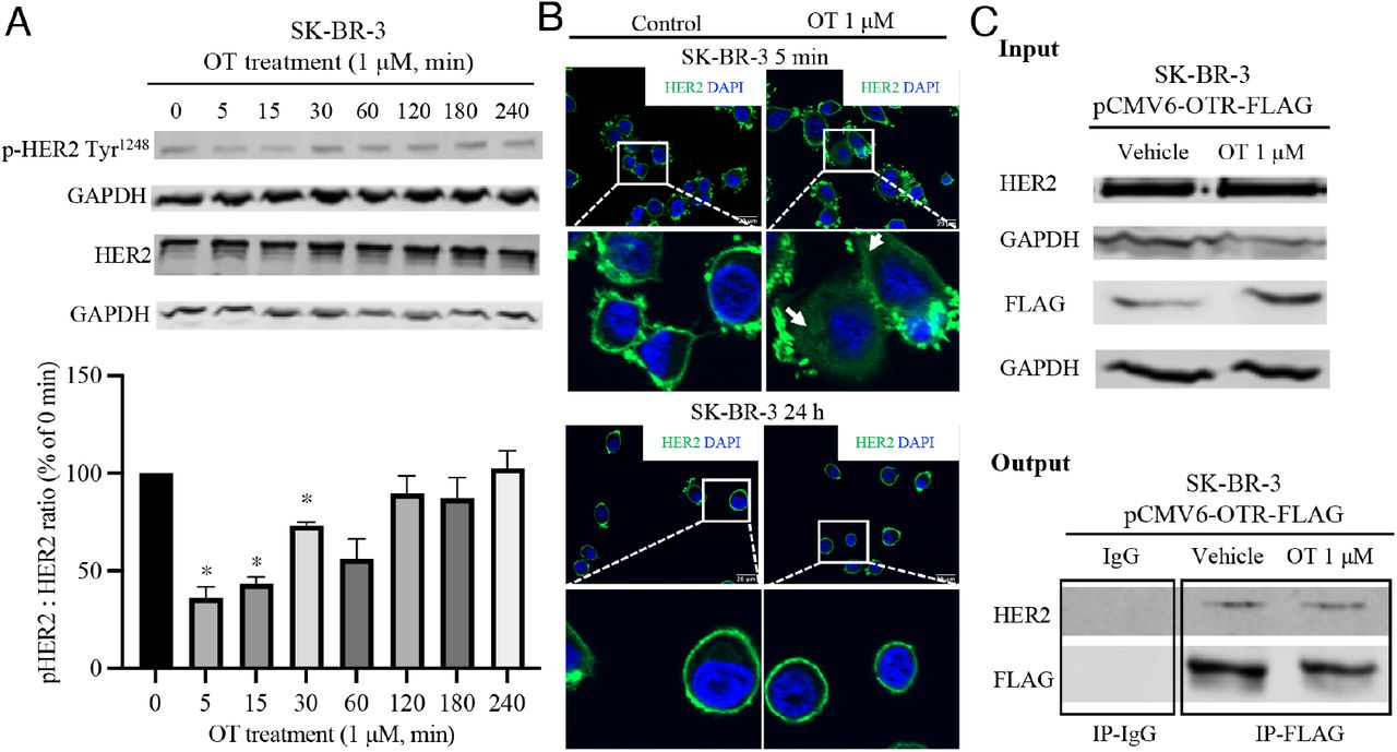
Journal: bioRxiv
Article Title: Oxytocin receptor and HER2 interactions in breast cancer
doi: 10.1101/2025.03.09.642136
Figure Lengend Snippet: A . Time course of HER2 phosphorylation in response to OT (1 μM) treatment detected by western blot. The bar graph represents the quantification of the phosphorylation indicated by the pHER2:HER2 ratio. One-way ANOVA analysis followed by Tukey’s multiple comparisons test was used for statistical analysis. *, p < 0.05 vs. 0 min group without OT treatment; n ≥ 3. B . Confocal microscope images of immunofluorescence for HER2 (green) with DAPI nucleus staining (blue) in SK-BR-3 cells treated with OT (1 μM) for 5 min and 24 h. HER2 was visualised with a primary rabbit antibody that specifically binds to the HER2 protein, followed by an Alexa Fluor 488-conjugated goat anti-rabbit secondary antibody using a Fluoro 2 – Zeiss Apotome 2.0 confocal microscope. The magnification for the top panel images is 630× (scale bar 20 μm), and the bottom panel images represent enlargements of the boxed areas from the top panel. Control, cells without OT treatment. C . Co-immunoprecipitation (Co-IP) demonstrated interaction of OTR with HER2. Total protein extracts from the cell lysis (input) were immunoprecipitated with anti-FLAG antibody (targeting the OTR-FLAG) and the mouse IgG (negative control) and resolved on SDS-PAGE gel. Protein interaction was immunodetected using HER2 and FLAG antibodies. Heterodimer of HER2 and OTR-FLAG was observed (output).
Article Snippet: The membrane was probed with the following primary antibodies: mouse-antibody for DYKDDDDK tag (FLAG) (MA191878, Invitrogen, Rockford, IL, USA), rabbit anti-HER2 (2165; Cell Signaling Technology, Beverly, MA, USA),
Techniques: Western Blot, Microscopy, Immunofluorescence, Staining, Control, Immunoprecipitation, Co-Immunoprecipitation Assay, Lysis, Negative Control, SDS Page

Journal: bioRxiv
Article Title: Oxytocin receptor and HER2 interactions in breast cancer
doi: 10.1101/2025.03.09.642136
Figure Lengend Snippet: A . Proposed model of HER2-OTR internalisation. OTR activation induces ERK1/2 and Akt phosphorylation. HER2-mediated signalling is either initiated by homodimerization when HER2 is expressed at high levels or by ligand-induced heterodimerization with other HER family receptors (HER1/HER3/HER4). Receptor dimerization causes transphosphorylation of tyrosine residues within the intracellular domains of the HER family receptors. Phosphorylation of HER2 reveals its binding sites for adaptor or effector proteins such as Ras/Raf and PI3K, which further activate ERK and Akt pathways, promoting cell survival and proliferation. B & C . Representative western blot images of total and phosphorylated ERK1/2 and Akt induced by OT treatment (1 μM). Column figures represent the quantification of the phosphorylation indicated by pERK1/2:ERK1/2 or pAkt:Akt ratio compared with 0 min group (n = 3 independent experiments). One-way ANOVA analysis followed by Tukey’s multiple comparisons test was used for statistical analysis. *, p < 0.05, **, p < 0.001 vs 0 min group (without OT treatment).
Article Snippet: The membrane was probed with the following primary antibodies: mouse-antibody for DYKDDDDK tag (FLAG) (MA191878, Invitrogen, Rockford, IL, USA), rabbit anti-HER2 (2165; Cell Signaling Technology, Beverly, MA, USA),
Techniques: Activation Assay, Binding Assay, Western Blot
Journal: Journal of Biological Chemistry
Article Title: Hyaluronan Constitutively Regulates Activation of COX-2-mediated Cell Survival Activity in Intestinal Epithelial and Colon Carcinoma Cells
doi: 10.1074/jbc.m703811200
Figure Lengend Snippet: FIGURE 1. Increased synthesis of HA stimulates cell proliferation, nuclear re-distribution of -catenin, and activation of cell survival proteins in HIEC6 human intestinal epithelial cells. A, HIEC6 cells infected overnight with a recombinant HAS2 adenovirus (HIEC6-HAS2-Ad cells) or a control -galactosidase adenovirus (HIEC6--gal-Ad cells) were washed, seeded (5,000 cells/well) in a 96-well cell culture plate, incubated for 72 h, and then assayed for cell proliferation by measuring mitochondrial enzyme activity (Celltiter 96R Aqueous One Solution cell proliferation assay kit (Promega)). Results are presented as mean S.D. of three independent experiments. B, control HIEC6--gal-Ad cells formed well organized epithelial clusters with tightly associated borders and peripheral -catenin. The HAS2 adenovirus- infected cells became scattered and spindle-shaped, and -catenin was dis- persed and translocated to the nucleus potentially reflecting greatly reduced -catenin-E-cadherin complex in the membrane as found in EMT. C, cell lysates from -galactosidase or HAS2-overexpressing HIEC6 cells were pro- cessed for immunoblotting for phospho (p)-ERK1/2, total (T)-ERK1/2, p-ErbB2, T-ErbB2, p-AKT, and T-AKT. Overexpression of HAS2 stimulates p-ERK1/2, p-AKT, and p-ErbB2 (2–4-fold). No significant changes in the levels of T-ERK, T-AKT, and T-ErbB2 were observed. Results in B and C are representative of three independent experiments.
Article Snippet: Rabbit polyclonal antibodies against ErbB2 and
Techniques: Activation Assay, Infection, Recombinant, Control, Cell Culture, Incubation, Activity Assay, Proliferation Assay, Membrane, Western Blot, Over Expression

Journal: Journal of Biological Chemistry
Article Title: Hyaluronan Constitutively Regulates Activation of COX-2-mediated Cell Survival Activity in Intestinal Epithelial and Colon Carcinoma Cells
doi: 10.1074/jbc.m703811200
Figure Lengend Snippet: FIGURE 5. HA regulates ErbB2 and AKT phosphorylation and -catenin expression, and this activation is reversed by HA-CD44 interaction antagonists. A, HIEC6 cells infected overnight with recombinant HAS2-Ad (HIEC6-HAS2-Ad) or -gal-Ad (HIEC6--gal-Ad as control) were incubated with or without CD44 siRNA (100 pmol) (control, scrambled siRNA) or O-HA (100 g/ml). The cells were lysed and processed for Western blotting for COX-2, phospho-(p-)AKT, total (T-) AKT, p-ErbB2, T-ErbB2, -catenin, and -actin (loading control). HIEC6- HAS2-AdcellsshowedincreasedCOX-2,p-AKT,p-ErbB2,and-cateninlevels.IncubationofthecellswithCD44 siRNA reversed the increase in these proteins by 80–90%. O-HA has a similar effect as CD44 siRNA. B, HCA7 cells were treated with 2 M AG825, infected with DN-AKT-Ad, or transfected with CD44 siRNA. Following treatment for 48 h, lysates were prepared and processed for Western blotting as in A. Treatment with CD44siRNA, DN- AKT-Ad, or AG825 (2 M) inhibited COX-2 and -catenin expression as well as p-AKT and p-ErbB2. C and D, densitometric quantitation of Western blot results from A and B are normalized. Results in A and B are repre- sentative of three independent experiments. Results in C and D are presented as mean S.D. of these three experiments. Variability in the expression of p-ErbB2, p-AKT, COX-2, and -catenin under each of the various conditions in these experiments is 4–10%. The differences in expression of these proteins with respect to the specific control are statistically significant (p 0.001).
Article Snippet: Rabbit polyclonal antibodies against ErbB2 and
Techniques: Phospho-proteomics, Expressing, Activation Assay, Infection, Recombinant, Control, Incubation, Western Blot, Transfection, Quantitation Assay

Journal: Journal of Biological Chemistry
Article Title: Hyaluronan Constitutively Regulates Activation of COX-2-mediated Cell Survival Activity in Intestinal Epithelial and Colon Carcinoma Cells
doi: 10.1074/jbc.m703811200
Figure Lengend Snippet: FIGURE 6. HA, ErbB2, PI3K/AKT, and -catenin regulate COX-2 expression. A, effects of -catenin siRNA transfection on COX-2 expression in HCA7 cells. Lanes 1 and 2, Western blots of lysates of HCA7 cells transiently transfected with control siRNA or pUSEamp empty vector control, respectively; lanes 3 and 4, transiently trans- fected with -catenin siRNA without or with co-transfection with CA-PI3K respectively; lane 5, transiently transfected with CA-PI3K. -Actin was a loading control. -Catenin siRNA down-regulated COX-2 and, as expected, -catenin expression. These inhibitions were not restored by co-transfection with CA-PI3K. B, effects of DN-PI3K transfection on COX-2 expression in HCA7 cells. Lanes 1 and 2, respectively, Western blots of empty vector pUSEamp- or pSV2-transfected HCA7 cell lysates (controls); lanes 3 and 4, HCA7 cells transiently trans- fected with pUSEamp-DN-PI3K without or with co-transfection with CA-ErbB2, respectively; lane 5, HCA7 cells transiently transfected with CA-ErbB2. -Tubulin was a loading control. DN-PI3K inhibited COX-2, -catenin, p-AKT, p-ErbB2 expression, and these inhibitions were not restored by co-transfection with CA-ErbB2. How- ever, CA-ErbB2 transfection alone increased p-ErbB2 and p-AKT expression over the endogenous levels of these proteins but has little effect on endogenous -catenin and COX-2 expression in HCA7 cells. Results presented are representative of three experiments for A and B. C and D, the densitometric values for Western blots of COX-2, p-AKT, p-ErbB2, and -catenin were normalized with respect to the -actin (C) and -tubulin (D) as loading controls. Results are presented as mean S.D. of three independent experiments. Variability in expression of p-ErbB2, p-AKT, COX-2, and -catenin under each of the various conditions in these experiments is 4–10%. The differences in expression of these proteins with respect to the specific control are statistically significant (p 0.001).
Article Snippet: Rabbit polyclonal antibodies against ErbB2 and
Techniques: Expressing, Transfection, Western Blot, Control, Plasmid Preparation, Cotransfection

Journal: Journal of Biological Chemistry
Article Title: Hyaluronan Constitutively Regulates Activation of COX-2-mediated Cell Survival Activity in Intestinal Epithelial and Colon Carcinoma Cells
doi: 10.1074/jbc.m703811200
Figure Lengend Snippet: FIGURE 8. Hyaluronan-COX-2-mediated effects on colon cancer cell sur- vival. In this study we have shown that perturbing constitutive HA-CD44 interactionbyHAoligomersandCD44siRNAdecreasesCOX-2expressionand enzyme activity by inhibiting an ErbB2 3 PI3K/AKT 3 -catenin 3 COX-2 axis and downstream anti-apoptotic events that contribute to cell survival. However, inhibition of COX-2 also decreases HA production suggesting the possibility of an amplifying, positive feedback loop between HA and COX-2. The possible feedback loop is represented by a dotted line in the figure.
Article Snippet: Rabbit polyclonal antibodies against ErbB2 and
Techniques: Activity Assay, Inhibition